14 Nov 2018

Using Mini-Genomes to Study Deadly Diseases


One of the major problems faced by scientists when studying contagious diseases is the threat of scientists themselves contracting the studied disease. In some cases researchers work in specialised labs with high security and lots of personal protection equipment. However, this is not always practical option. I recently spoke to Rebekah Penrice-Randal, a 1st year PhD student in IGH, about her project on Ebola and how she is using mini-genomes to study them without the risk of infection.

First, what is Ebola? Ebola (also known as Ebola Virus Disease or Ebola Hemorrhagic Fever) is a viral disease that has been in the news in the past few years, especially 2014-16 mainly due to a major outbreak that occurred in west and central Africa. According to the World Health Organisation (WHO) there were around 28,000 cases and over 11,000 deaths and there is currently no licenced vaccine for this disease.

Patients who contract Ebola deteriorate very quickly. Within 2-21 days of becoming infected sufferers can start with symptoms including: fever; headache; muscle weakness and a sore throat. These often progress to vomiting and diarrhoea, stomach pain and unexplained bleeding (haemorrhages).
The disease is spread through direct contact: through broken skin; contaminated bodily fluids; contaminated needles. Ebola can also be contracted from infected animals such as bats, apes or monkeys. The virus often remains undetected by the immune system in certain bodily fluids e.g. semen, breast milk, ocular (eye) fluid and spinal column fluid even after someone has recovered.

So now we know what Ebola is, what are mini genomes and how do they help in the study of this disease?

Viruses have genes, some of which allow them to replicate or make more of themselves when they are within host cells. These genes have a start and stop regulatory sequence which tells the cell machinery that transcribes the genes where the gene starts and where it finishes(Transcription is the process by which the genes are converted into messenger RNA, an intermediated step which is then converted into proteins). A mini-genome is a shorter version of the Ebola genome. The regulatory sequences are kept but the viral genes in-between that make the virus ‘infective’ are removed and replaced with a reporter gene. A reporter gene is simply a gene which has easily identifiable and selectable markers, one such example is green fluorescent protein (GFP), naturally produced in jellyfish which, when expressed, fluoresces in green. The amount of fluorescence can then be easily measured.

A change in the environment can affect the regulatory sequences and in turn the amount of protein that is produced and measured. These mini genomes can be inserted into a bacterium like E. coli and when environmental conditions are changed the effects to the mini genome genes can be seen. This means you can study the transcription and replication of these genes safely and without the dangerous viral genes being produced.

Rebekah is also going to compare the transcriptomes i.e. the measure of all genes that are expressed at a particular time, both of the mini genome infected cells and the actual Ebola infected cells. This will allow her to see how representative the mini genomes are as a model. This can also show how the disease can differ depending where in the world a sample is collected and can show natural variation within the virus. Furthermore, when the environmental conditions are changed, such as by reducing the amount of oxygen, the evolution of the virus under those conditions can also be seen.

This research will contribute in the quest to understand and put an end to this deadly disease.

Eleanor Senior is a 3rd Year PhD student in IGH studying the bovine parasite Tritrichomonas foetus.

11 Oct 2018

From Snails to Sheep: The Flatworm Affecting Farms Across The UK


Farm animals are susceptible to a whole range of diseases and parasitic infections. From the commonly known and well publicised foot and mouth disease to the lesser publicly recognised bluetongue disease, farmers must deal with a wide array of viruses, bacteria and parasites that can affect their livestock. In this blog, I spoke to Bethan John, a 3rd Year PhD student in IGH about her PhD research into Liver Fluke.

Liver fluke (Fasciola hepatica) is a flatworm parasite of grazing animals such as cows and sheep. Though only the size of a 50p piece, this parasite causes weight loss and anaemia in infected animals and is estimated to cost the UK cattle industry £40.4 million per year. Therefore, it is both an animal welfare issue and an economic one. This parasite is also known to infect humans (zoonotic) when humans eat infected meat.

The liver fluke takes many forms during its life cycle. Infection occurs when the animals consume the parasite when it is in the form of a cyst attached to blades of grass. Once the cysts are in the small intestine they mature into juvenile parasites which burrow from the small intestine across the peritoneum and into the liver where they mature into adults. These adults are hermaphrodites having both male and female characteristics, and are able to reproduce both sexually (2 parents) and asexually (1 parent) resulting in the production of eggs.

These eggs are passed from the mammal via its faeces onto damp pastures where they hatch. They are then able to infect the mud snail (usually Galba truncatula) which is the intermediary host. Within the snail the parasite  is cloned into thousands of genetically identical cysts, a process known as clonal amplification. It is these genetically identical cysts  that are shed and become attached to blades of grass. This process can only occur in boggy areas where the snail can be found. The cycle is then completed when the parasite cysts are ingested by grazing animals and mature flukes develop in the liver and go on to lay eggs.

However, there has been evidence via word-of-mouth that even animals that have been housed inside and fed on silage, far away from boggy areas and snails, have still become  infected with fluke. Silage, which is grass that has been dried and fermented in airtight conditions, has also been known to transmit some bacterial diseases and other parasites. It is, therefore, thought that the fluke cysts could be transmitted from the silage to the housed animals. Moreover, it may also be the case that the eggs can survive in slurry, animal waste mixed with water and runoff, which is commonly used as a fertiliser on farms.

As part of her PhD Bethan is looking into whether the various fluke life cycle stages  can survive  under certain environmental conditions and within silage  Fluke eggs are a lot more sensitive to the environment than the cysts, they need moisture and a temperature over 10°C to hatch, whereas cysts can survive in much cooler environments; over 50% of cysts on grass can overwinter and still be infectious to grazing animals.

There is also a big issue with the use of  drugs to control fluke infections. The usual drug used, triclabendazole, is becoming less effective. If it can be shown that fluke can be passed to livestock via silage then farmers can change and improve their farming practises to reduce the incidence of fluke without the need for drugs.

Eleanor Senior is a 3rd Year PhD student in IGH studying the bovine parasite Tritrichomonas foetus.

                                          Bethan hunting for snails on a UK farm.


11 Sept 2018

The cow STI costing America over $200 million


Eleanor Senior is a 3rd Year PhD student at the Institute of Infection and Global Health. Here she shares her research investigating vaccine candidates for bovine sexually transmitted infection.

Many types of animal parasite are very well known for example, most people have heard of things like fleas, tapeworms and ticks. However,  there are many thousands of parasites that exist that you may never have heard of, but are no less important. Once such parasite is Tritrichomonas foetus.
This is a microscopic parasite that is sexually transmitted between cows and bulls causing a disease called Bovine Trichomoniasis, which is related to the human STI Trichomoniasis, caused by Trichomonas vaginalis.

T. foetus is found in many countries across the world including the USA, Brazil, France, Germany and Australia. Fortunately, this parasite does not infect cattle in the UK because of this country's strict border controls and the use of artificial insemination (AI); a procedure that allows for screening of sperm for infections prior to insemination.

So what is so bad about Bovine Trichomoniasis anyway? This parasite often doesn’t cause any symptoms in bulls in the same way that many STIs are symptomless in men. Consequently, it is difficult to detect in bulls unless there is constant sperm testing. Likewise, it is also difficult to spot in the cows.  Often it causes early stage spontaneous miscarriage which can be so early that the farmer isn't aware that the cow is pregnant in the first place. It can also cause short or long-term infertility. The first indication for the farmer that the herd is infected is that there is a decrease in the number of cows becoming pregnant or carrying to full term. Again, the best way to find out whether the parasite is present in the stock is to test all the cows. This can be pretty tricky and time consuming, particularly when there could be thousands of cows on a farm or ranch which will need to be tested at regular intervals.

The lowered calving rates and lower levels of milk production as a consequence of this parasite infection has significant economic consequences for the farmer.  Furthermore, the need for the infected cattle to be destroyed as a means for protecting the rest of the herd takes an additional financial toll.  It has been estimated that in Texas alone the losses due to this parasite can reach $195 million. Taking into account the losses from other states in America the amount is a considerably higher. So this parasite is clearly a problem both financially for the farmers and as a welfare issue for the cattle.

Although there are currently vaccines available to combat Tritrichomonas foetus, there are none as yet that prevent reinfection. Consequently available vaccines must be given at each breeding season. This again isn’t very practical when there can be thousands of cows to immunise every month or two.
My PhD is aiming to find proteins from the parasite itself that can be used as vaccine candidates. We are searching for a small group of proteins that we can further test for key vaccine qualities such as:
  • promoting an immune response
  • being found in all variants of the parasite across the globe not just in one region. This is so that one vaccine can be used against all variants rather than multiple vaccines each of which deals only with one or two specific variants
To do this I am using a reverse vaccinology approach. So what is this? Classical vaccinology is what has been used to make lots of the common vaccines available today such as the flu vaccine. In these vaccines, weakened or killed viruses or bacteria are used to promote an immune response. However, the exact protein or proteins that causes this response are often not known. Reverse vaccinology involves using computers to predict proteins that are likely to give an immune response which are then narrowed down using a variety of laboratory techniques to give a small number of candidates. Some, or all, of these candidates are then used as the starting point to produce the actual vaccine.

By the end of my PhD I hope to have come up with a list of potential proteins that can then be used to start designing a vaccine in the fight against this costly and distressing parasite.


23 Mar 2018

How to help people with tuberculosis avoid the medical poverty trap


File 20180321 165580 i4eyto.jpg?ixlib=rb 1.1
Shantytown near Lima, Peru. Inspired By Maps/Shutterstock.com
Tom Wingfield, University of Liverpool 
Rosario hacks into a handkerchief, coughing up the bloodstained phlegm that plagues her chest in the mornings. On hearing the noise, two heads pop up from under a blanket – Rosario’s twins, Gonzalo and Bruno. A wail starts up from the crib in the corner of the one-room shack. As she puts baby Angelita to her breast, Rosario ponders her situation.

It’s been half a year since her husband Samuel passed away, two months since this horrible cough started, and six days since the doctor told her she had tuberculosis and she started the medicines. Nearly every day since then, Rosario has had to make the hour-long, bumpy minibus journey to the tuberculosis clinic – Angelita in hand – so the nurse can witness her taking the drugs.
But Rosario just can’t get to the clinic today. Her neighbour can’t look after the twins, and Rosario has no money to pay for the minibus ride. More pressingly, there is no food left to cook nor kerosene to cook with.

Rosario quickly calculates that, after deducting two packets to feed the twins, she has enough biscuits left to sell for a small profit at the minibus stop. For today, at least, that will stop the family going hungry. But it will also mean not arriving at the clinic until after it shuts – too late to take her medicines.

A social disease

Rosario’s situation in a Peruvian shantytown is not fictional, not isolated, and not new. Nearly a century and a half ago, Rudolf Virchow, the father of social medicine, recognised that tuberculosis (TB) and poverty were inseparably linked in a vicious cycle. He called TB “a social disease”. Indeed, the improvements in poverty levels, living conditions and nutrition that occurred during the Industrial Revolution in Europe were associated with a fall in TB rates, many years before the discovery of the TB bacteria or TB medicines. Today, the poorest households continue to suffer the highest levels of infectious diseases and, in trying to access healthcare, can be pushed deeper into poverty and ill health – the so-called “medical poverty trap”. There is no disease that better typifies this trap than TB.

Rudolf Virchow, the father of social medicine. Wikimedia Commons

Despite this, recent global TB control strategy has been disproportionately focused on medicines and tests rather than addressing the social causes of the TB epidemic. And so, today, Rosario’s terrible dilemma continues to be faced by many of the roughly 10m people worldwide who will develop tuberculosis this year (1.3m of whom will die).

This is one of the reasons why the global response to TB is not working. A more holistic approach to TB control is needed that addresses not just the disease but also the person who has the disease and the circumstances in which they live.

In its 2015 End TB Strategy, the World Health Organisation (WHO), for the first time in the modern era of TB control, called for social support and poverty alleviation strategies for people with TB to reduce the hidden costs of treatment, reduce stigma, empower patients, and increase TB prevention, the number cured and their overall well-being. But evidence that this type of strategy works was limited.

Testing the theory in Peru

The multi-disciplinary Innovation For Health and Development research team, which I joined in 2010, has been working for the past two decades in shantytowns near Lima, Peru, to generate new evidence to fill this knowledge gap and support TB-affected households.
One of our first tasks was to measure households’ hidden costs of TB treatment. Hidden costs (like those Rosario faces) included travel to clinics, food and lost income.

We found that when these hidden costs exceeded a fifth of a household’s annual income, the patient in the household was more likely to abandon treatment, fail treatment or die. In essence, the threshold of costs that we had measured had been catastrophic, not only to household finances but also to the TB patients’ health. This threshold, among others, was subsequently endorsed by WHO in their TB patient costs survey, which is being deployed around the world.

Our findings had identified a crucial factor explaining why medicines alone were not controlling TB. In response, we provided social and financial support for TB-affected households. Financial support consisted of bank transfers (up to US$40 per month) to reduce the hidden costs of TB and enable access to TB care. Social support included household visits by our research nurses and mentoring from mutual support groups led by former TB patients to empower households to access care and reduce stigma.

The intervention was a success. Supported TB patients were significantly more likely to complete their treatment or be cured, and their children were more likely to take medicine to prevent TB.
Rosario (not her real name) was one of the patients who received this support. Using the money she received throughout her treatment, she was able to keep going to the clinic and ensure that Gonzalo, Bruno and Angelita were fed regularly, and took medicine to prevent TB. She is now cured of TB and able to work.

The ConversationHelping households affected by TB to avoid the medical poverty trap, and providing them with moral support and hope, can enhance TB care and prevention. Without it, we won’t achieve the End TB Strategy goal of eliminating the disease by 2050, and millions more vulnerable households, like Rosario’s, could continue to suffer an entirely avoidable downward spiral of poverty and ill health.
Tom Wingfield, NIHR Academic Clinical Lecturer in Infection and LIV-TB Collaboration Lead, University of Liverpool

This article was originally published on The Conversation. Read the original article.

14 Mar 2018

Five diseases you can catch from pets

File 20180312 30972 vw8rut.jpg?ixlib=rb 1.1


Love them or hate them, it is hard to get away from pets. And even if you don’t own one yourself, you are likely to come across them (or things they have left behind) regularly.

Most interactions between humans and pets are likely to be overwhelmingly positive. But pets can carry some diseases that affect us. Such diseases, termed zoonoses, are usually very mild, but the rarer ones can be more severe.

Here are some of the infections people can catch from their pets:

1. Rabies

Rabies is perhaps the archetypal zoonosis. A virus whose name alone has the potential to cause fear. The virus is largely found in unvaccinated dogs and other canine populations.

In areas that still have rabies, people – often children – usually become infected when they are bitten by an affected dog. The virus attacks the brain, and once symptoms develop, there is sadly no cure, and those affected die. The good news is, it can be prevented by vaccinating dogs and other wild carnivores. Many parts of the world are now free of the virus, including the UK and large parts of the rest of Europe, and in many others, national campaigns are under way to achieve this.

2. Ringworm

Some zoonotic skin infections are not uncommon in pets but usually mild in humans. These can be shared with owners because of our love for warm houses, and close contact with our pets. Ringworm is one such infection.

Ringworm is actually a misnomer. It is not a worm at all but a microscopic fungus, closely related to the cause of athlete’s foot in people. Affected cats, dogs and other animals may show very few signs. However, in its classical form, pets with ringworm usually have circular areas of hair loss. The affected area of skin becomes scaly, flaky and itchy. It is very treatable, but can occasionally cause scarring.

3. Salmonella

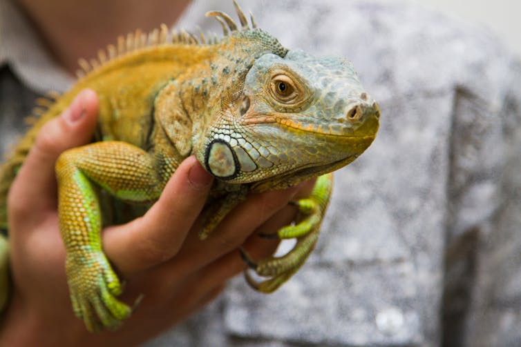
A variety of potentially zoonotic bugs live in the intestines of pets. These rarely affect humans. However, when they do, they can be severe. We have all probably heard of salmonella, largely because of risks, now thankfully much diminished, from eggs. Dogs and cats can also carry salmonella, sometimes causing diarrhoea. Salmonella is also quite commonly present in pet reptiles and amphibians, as well as in so-called “feeder mice” that are fed by some to pet reptiles.
It’s always a good idea to wash your hands after handling both pets and raw pet food. It is also a good idea to have separate areas for preparing raw animal food and human food.


Pet reptiles can carry salmonella. SGr/Shutterstock.com

4. Toxoplasma

Toxoplasma is a common parasite in cats that they can also shed in their faeces. For most humans, it is entirely benign. However, if a woman first becomes infected during pregnancy, it can, albeit rarely, have severe complications for the developing foetus.

Pregnant women should take simple additional precautions around hand hygiene, avoiding cat litter trays, especially those not cleaned regularly, and avoiding eating uncooked garden produce where cats may have had access to the soil.

5. Bites and scratches

Some argue for bites and scratches to be included as a zoonosis. If we do include them, they are likely to be among the most common zoonoses. Never nice, always painful, and in disturbing, rare cases – usually involving children – they can be fatal.

Cat bites and scratches can transmit a bacterium called Bartonella henselae, the cause of “cat-scratch disease”. Both bites and scratch wounds can become badly infected causing further pain. Scars, both mental and physical, can be lifelong in those that have been attacked. Children and those exposed occupationally, such as postmen, are perhaps most at risk.

As with most infections, zoonotic infections have a greater potential to do harm in people whose immune systems are compromised, such as the elderly and those suffering from immunosuppressive diseases (such as HIV/AIDS), or undergoing immunosuppressive therapies (such as chemotherapy). However, even if your immune system is compromised, you can still benefit from owning a pet. And with care and a little knowledge of the risks, you can avoid infections.

The ConversationThankfully, zoonotic infections are not common. Most infections we get are likely to come from other humans. However, the risks of zoonosis can be minimised by being aware of them and by taking simple hygiene precautions at home. And, if in doubt about the risks, you can always consult your GP or a veterinary surgeon.

Alan Radford, Professor of Veterinary Health Informatics, University of Liverpool
This article was originally published on The Conversation. Read the original article.
The Institute of Infection and Global Health. Powered by Blogger.